You know that moment when a kid asks "how do my eyes actually work?" and you suddenly realize you're not entirely sure yourself? Honestly, most of us fumble through a vague explanation about pupils and lenses, then quickly change the subject. But here's the thing: when you're handed a science worksheet label parts of the human eye, suddenly the stakes feel higher. It's not just about passing a quiz—it's about making that abstract diagram actually click for a restless 10-year-old who'd rather be on a tablet.
Look, I've been there. You print out that clean diagram of an eyeball, hand over a pencil, and watch them stare at "sclera" and "cornea" like they're ancient runes. The truth is, most worksheets fail because they treat the eye like a boring machine instead of the incredible, squishy camera it really is. But this matters right now because visual learning is how kids actually lock in concepts—and if the worksheet doesn't connect, neither will the lesson. You need something that makes them point to their own eye and say "oh, that's the iris!"
What I'm going to show you isn't just a list of terms to memorize. It's a way to turn that worksheet into a conversation starter—where the retina becomes a movie screen and the optic nerve is the high-speed cable. By the end, you'll have a method that makes labeling feel like detective work, not homework. And honestly? You might learn something new too. (I still can't believe the cornea has no blood vessels. Wild, right?)
Let's be honest about something: most anatomy worksheets teach kids to memorize parts in a vacuum. You label the cornea, the iris, the lens, and then you move on. But the human eye is not a static diagram. It's a living, fluid-driven optical instrument that adjusts itself faster than any camera on the market. When you hand a student a science worksheet label parts of the human eye, you're giving them a map of a machine that works on pressure, refraction, and split-second muscle reflexes. The real trick is making that map come alive.
Why the Lens Gets All the Attention (and the Retina Deserves More)
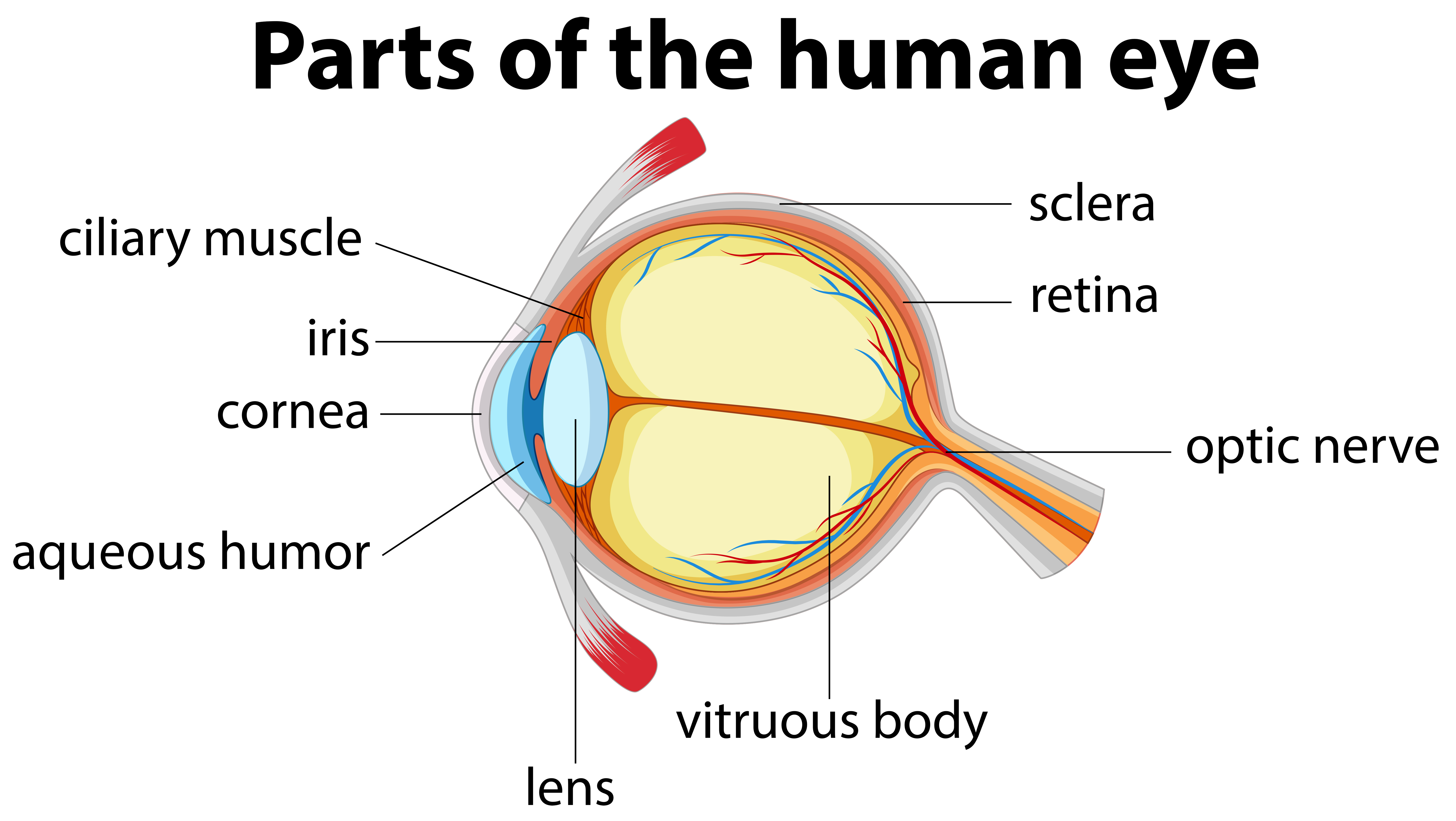
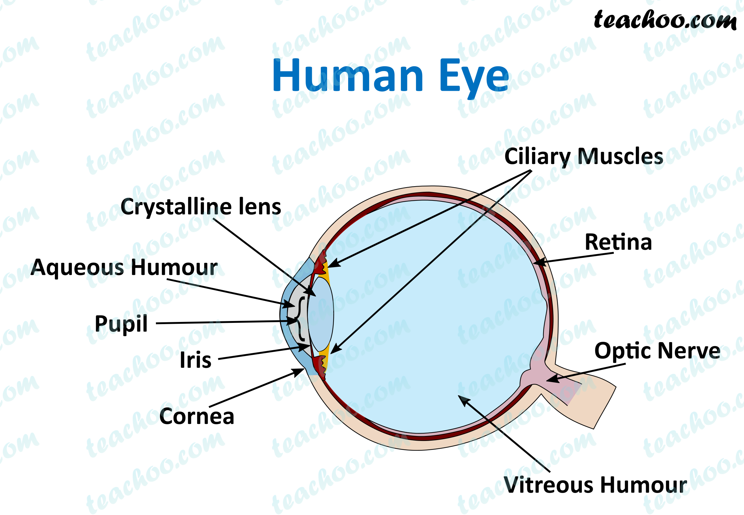
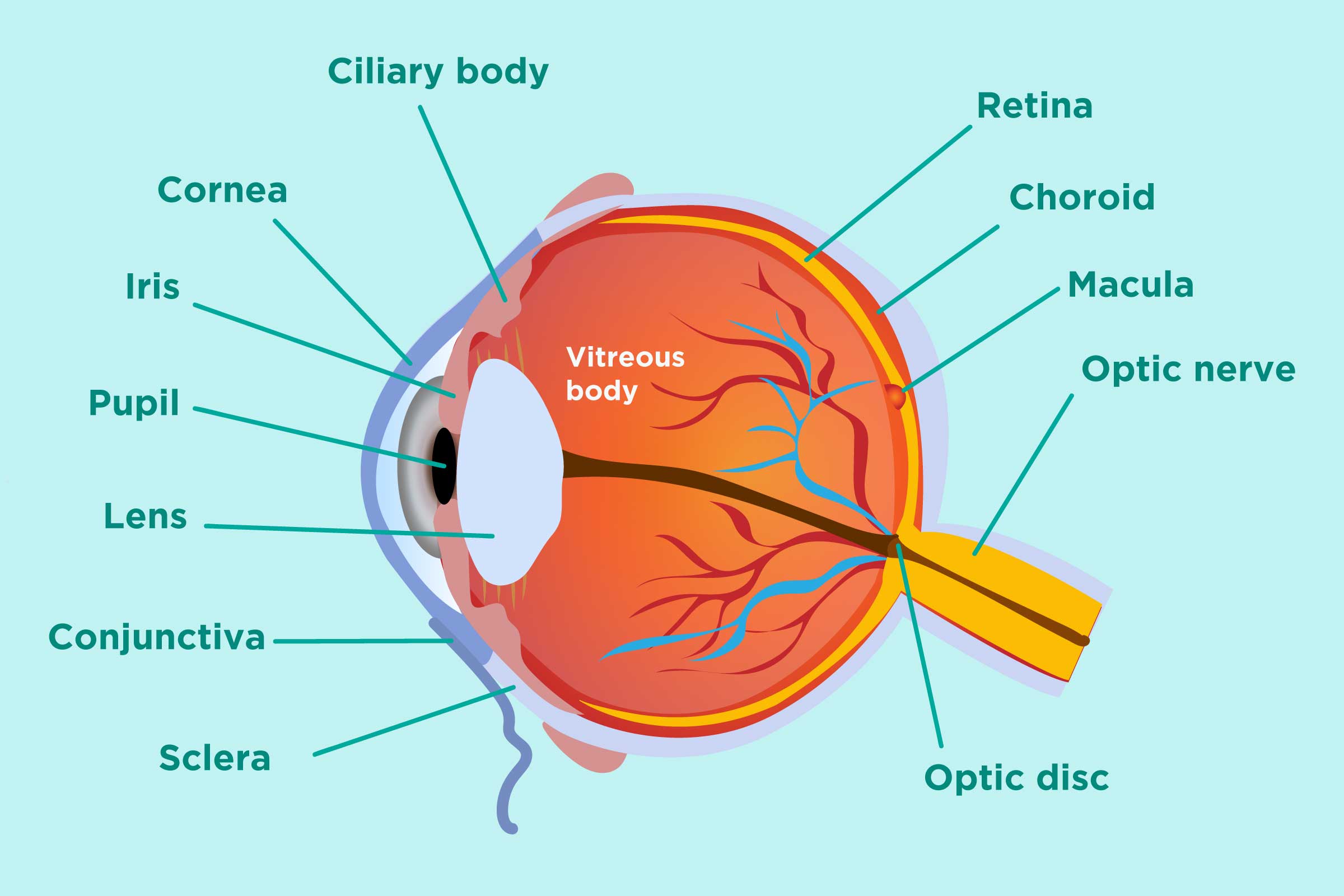
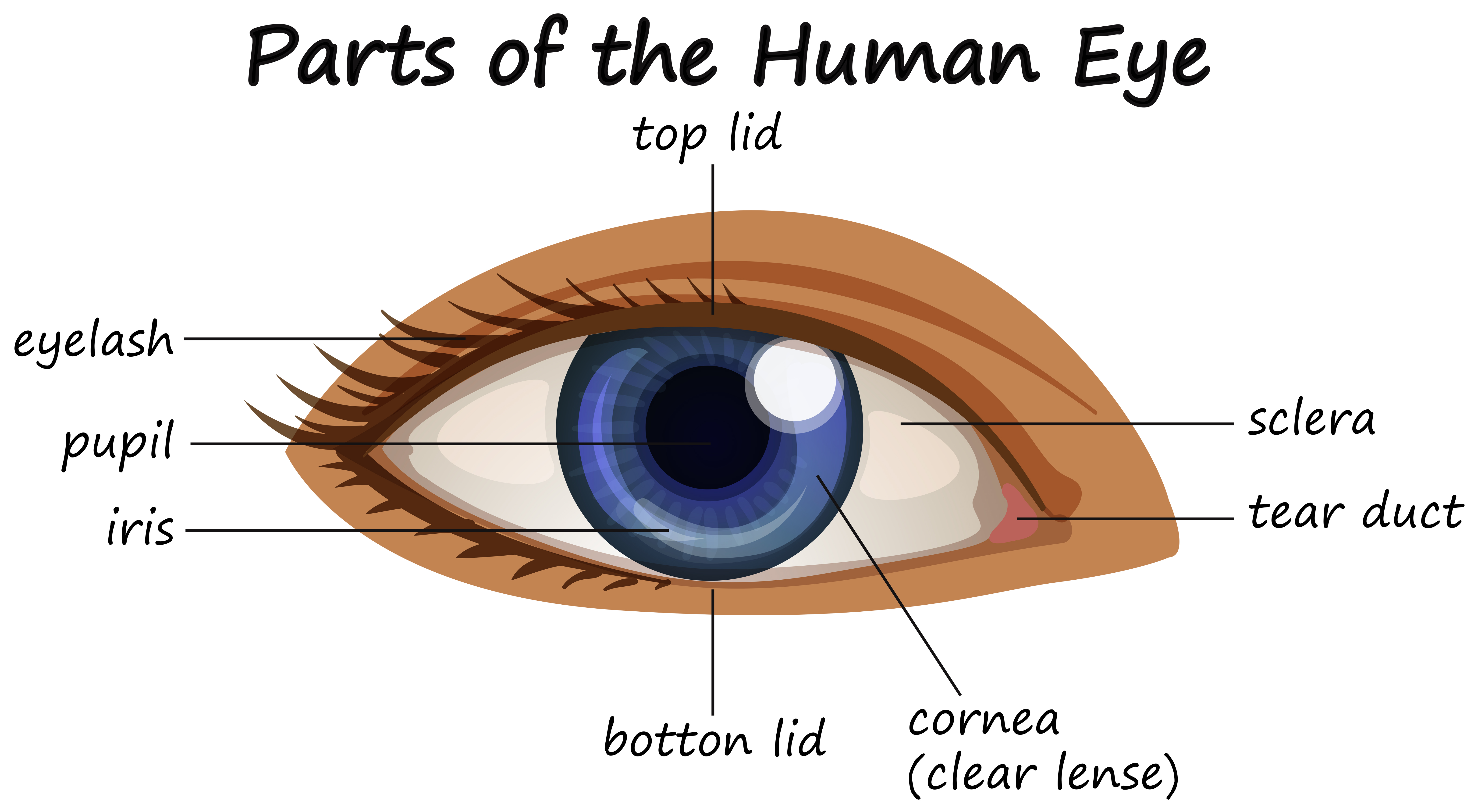
Every worksheet I've ever seen puts the lens front and center. It's the glamorous part, right? The lens changes shape, it focuses light, it's flexible. But here's what nobody tells you: the retina is where the actual magic happens. Without the retina, the lens is just a fancy piece of tissue projecting a blurry image onto a blank wall. The retina takes that inverted, upside-down image and converts it into electrical signals your brain can read. It's packed with photoreceptors—rods for low light, cones for color—and they're not evenly distributed. You have a blind spot where the optic nerve exits, and your brain literally fills in the gap. That's not science fiction; that's your daily reality.
When I teach this, I tell students to hold their thumb out at arm's length, close one eye, and slowly move the thumb sideways until it disappears. That's the blind spot in action. Suddenly, a diagram on a science worksheet label parts of the human eye becomes personal. It's no longer abstract anatomy; it's a demonstration of how your own vision is actively editing reality. That's the kind of sticky learning that lasts beyond Friday's quiz.
The Cornea Does the Heavy Lifting
Most people assume the lens does all the focusing. Wrong. The cornea—that transparent dome at the front—provides about two-thirds of the eye's total focusing power. It's fixed, it's curved, and it's the reason why LASIK surgery reshapes the cornea, not the lens. If the cornea gets scratched or swollen, your vision goes fuzzy immediately. The lens then fine-tunes that focus for near or far objects. Think of the cornea as the wide-angle lens on a camera and the lens as the autofocus mechanism. Neither works alone.
The Pupil and Iris: A Dynamic Duo
The iris isn't just for eye color. It's a muscular diaphragm that controls the pupil's size. Bright light? Pupil constricts. Dark room? Pupil dilates. This isn't passive—it's a reflex that protects your retina from damage. Here's a specific tip: shine a penlight in your own eye in a mirror. Watch the pupil snap shut. Then cover that eye for ten seconds and uncover it. The pupil will be smaller on the side that was covered because both pupils respond to light hitting either eye. That's called the consensual light reflex, and it's a real-world test neurologists use. Try that with a standard diagram and see if it holds up.
The Optic Nerve: The Forgotten Cable
Every worksheet labels the optic nerve, but few explain its vulnerability. This bundle of over a million nerve fibers has to exit the eyeball, creating that blind spot I mentioned earlier. But more importantly, increased pressure inside the eye can crush these fibers, leading to glaucoma. That's why eye doctors puff air into your eye during a checkup—they're measuring intraocular pressure. The optic nerve has no pain receptors, so damage happens silently. Knowing where it sits on a diagram might just remind someone why annual eye exams matter.
What Most Worksheets Get Wrong About How the Eye Actually Works
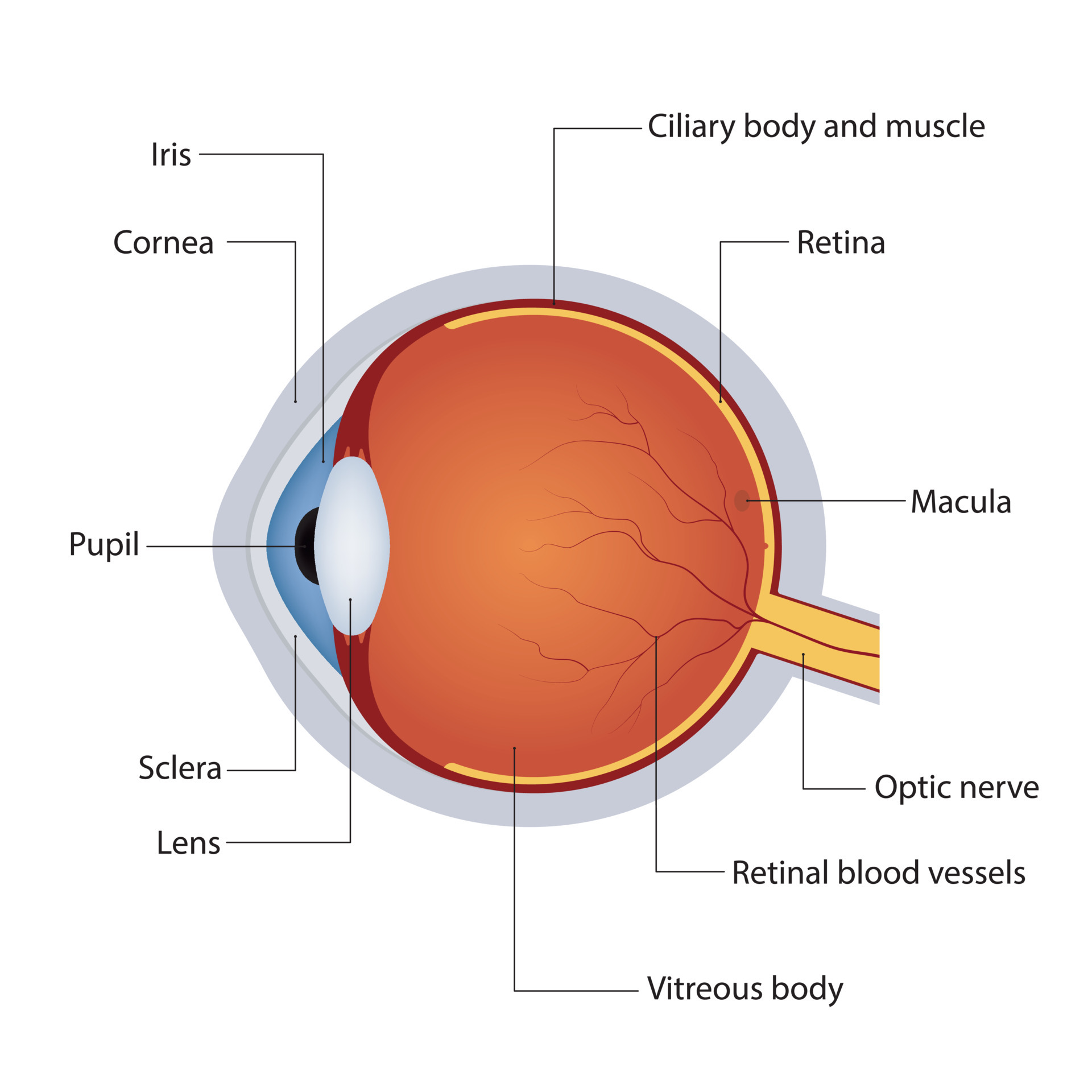
The classic diagram is usually two-dimensional and color-coded. It shows light entering straight through the center, hitting the fovea dead-on. Reality is messier. Light scatters, the eyeball is slightly asymmetrical, and the vitreous humor (that jelly filling the back of the eye) can have floaters. Plus, the image projected onto the retina is inverted and reversed left-to-right. Your brain flips it. That's not on any worksheet I've seen, but it's fundamental. If you're using a science worksheet label parts of the human eye, add a note in the margin: "The brain flips this image upside down." It changes everything.
Here's a quick breakdown of the three main refractive components and their real-world quirks:
| Structure | Primary Job | Common Misconception | Real-World Problem |
|---|---|---|---|
| Cornea | Provides ~65% of focusing power | People think the lens does all the work | Scratches cause immediate blur; LASIK reshapes this, not the lens |
| Lens | Fine-tunes focus (accommodation) | Believed to be the main focusing element | Cataracts cloud it; presbyopia stiffens it with age |
| Retina | Converts light into neural signals | Seen as passive "film" in a camera | Macular degeneration destroys central vision; retinal detachment is an emergency |
That table isn't just for memorization. It's a cheat sheet for understanding why an optometrist asks "better one or two?" during a refraction. They're testing how your cornea and lens work together to land the image precisely on your retina. Get that alignment wrong, and you get nearsightedness, farsightedness, or astigmatism. A good worksheet should lead to questions, not just answers. The best ones make you wonder why your own eyes work the way they do—and that curiosity is worth more than any labeled diagram.
Your Next Step Starts Here
Understanding how the eye works isn't just about passing a quiz—it's about appreciating the incredible machinery behind every glance, every smile you catch, every sunset you admire. Whether you're teaching a child, brushing up on your own biology, or helping a student connect the dots between anatomy and everyday life, this knowledge sticks when you actually use it. That’s where a simple tool like a science worksheet label parts of the human eye transforms abstract terms like "cornea" and "retina" into something real and memorable. It’s the difference between memorizing a list and truly understanding how you see the world.
Maybe you're thinking, But I'm not a science teacher—will this really help? The honest answer is yes. You don’t need a lab coat to make learning hands-on. A single worksheet, used with curiosity instead of pressure, can unlock a lightbulb moment for someone who’s been struggling. The hesitation you feel is just the gap between knowing you should do something and actually starting. Close that gap. Print it out, grab a pencil, and see where the process takes you.
Now that you’ve got the big picture, don’t let it fade. Bookmark this page so you can come back when you need a refresher, or share it with a fellow parent, tutor, or teacher who’s looking for that "aha" resource. And if you’re ready to explore more, browse the gallery of worksheets and diagrams here—each one designed to make learning feel less like work and more like discovery. Your next breakthrough is just one click away.